Paracolic Gutter Femoral Vein

Pin On Abdomen And Urogenital

The Peritoneal Cavity Greater Sac Lesser Sac Teachmeanatomy

Left Paracolic Gutter Radiology Reference Article Radiopaedia Org

Abdominal Wall Mesentery Peritoneum And Vessels Radiology Key

Subphrenic Spaces Medical School Stuff Abdominal Peritoneal Dialysis

Abdomen Mistryland
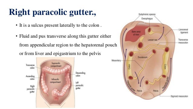
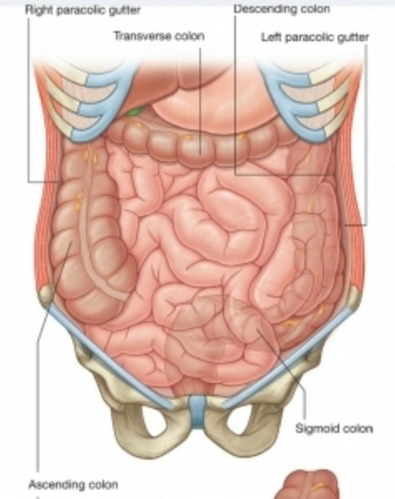
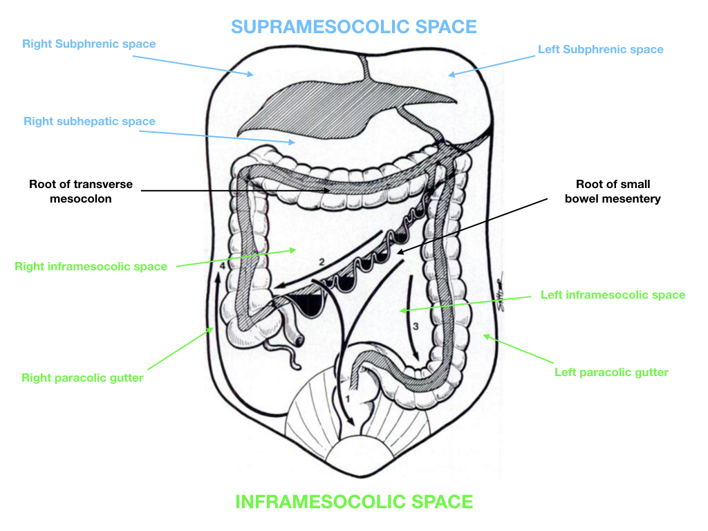
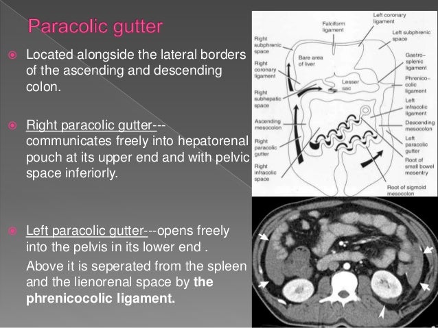
The right paracolic gutter is a component of the right inframesocolic space continuous superiorly with the right subhepatic and right subphrenic spaces it is larger than the left paracolic gutter which is partially separated from the left subphrenic spaces by the phrenicocolic ligament.
Paracolic gutter femoral vein. The left paracolic gutter is a component of the left inframesocolic space partially separated from the left subphrenic spaces by the phrenicocolic ligament. How may fluid get from the left paracolic gutter to the thorax. A less obvious medial paracolic gutter may be formed especially on the right side if the colon. The left medial paracolic gutter.
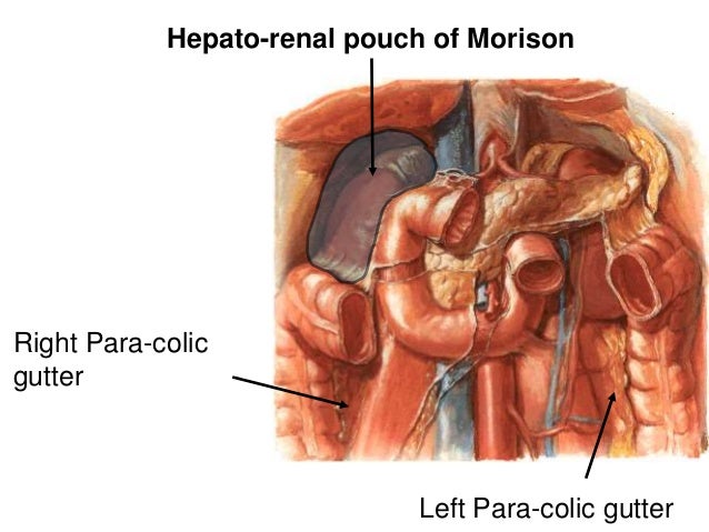
Its origin lies on the right side origin of the right paracolic gutter lies at the ascending portion of the colon at the right hepatic flexure or the point where the ascending colon turns at a right angle to form the transverse colon. Both paracolic spaces are in continuity with the pelvic peritoneal spaces. The right paracolic gutter is larger than the left and communicates freely with the right subphrenic space. It is smaller than the right paracolic gutter.
What vein connects the femoral vein with the axillary. Both paracolic spaces are in continuity with the pelvic peritoneal spaces. This is why this patient also has a hepatocellular carcinoma with cirrhosis ascites portal hypertension portacaval anastomosis and splenomegaly. Portal to portal or portal to caval.
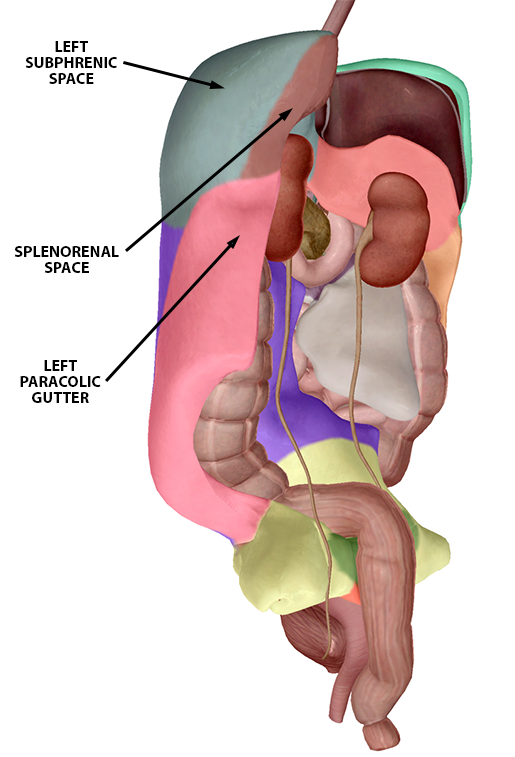
What makes up the portal triad. 2 ruq view hepatorenal space subphrenic space right paracolic gutter liver tip right thoracic cavity 3 luq view splenorenal space subphrenic space left paracolic gutter left thoracic cavity 4 pelvic view longitudinal and transverse view of the bladder. Hepatic artery proper portal vein bile duct. Due to back flow buildup in these veins creates this condition.
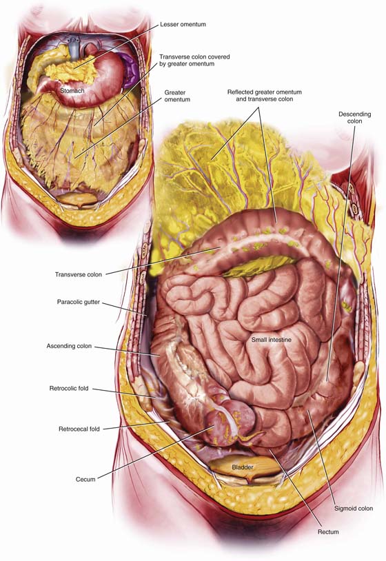
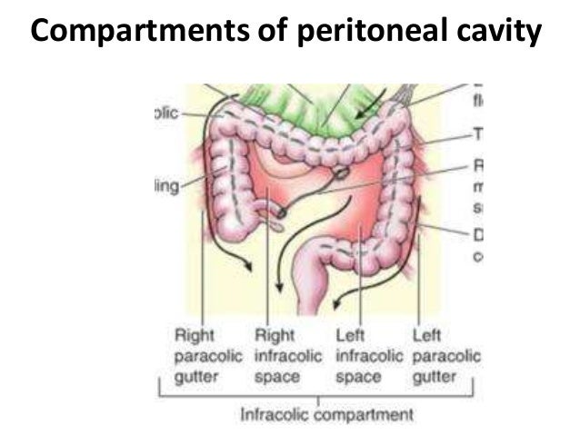
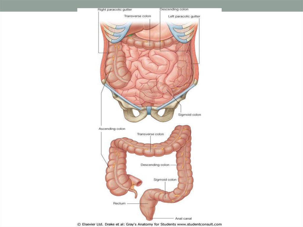
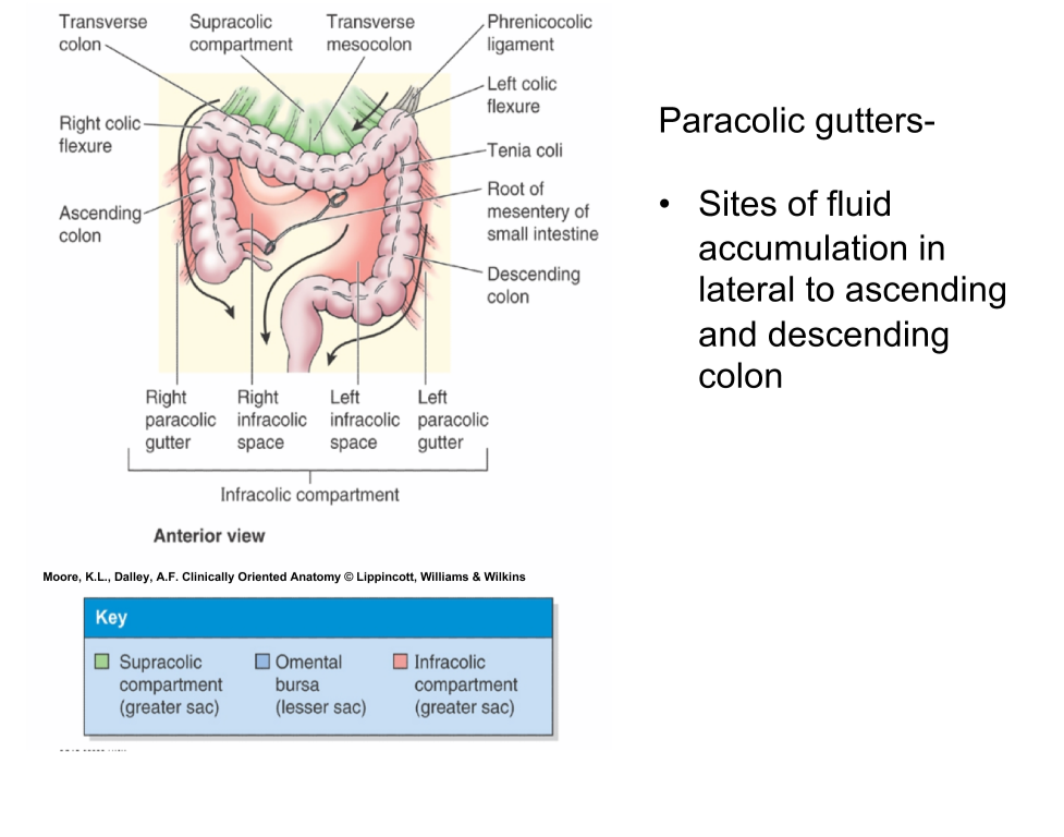
The main paracolic gutter lies lateral to the colon on each side. In the setting of abdominal pain whether acute or chronic ct is helpful and frequently essential in discovering the underlying cause fat stranding is a common finding on ct of the abdomen and when present it directs the radiologist s attention to the site of pathology. Gross anatomy origin posterior to inguinal ligament within lacuna vasorum 1 as continuation of femoral vein termination t. This allows the user to perfectly see the different parts of the peritoneal cavity omental bursa paracolic gutters mesentery mesocolon.
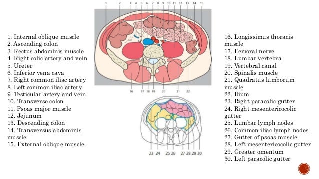
The paracolic spaces gutters are located lateral to the peritoneal reflections of the left and right sides of the colon fig 8a. The external iliac vein eiv is located along the pelvic brim between the inguinal ligament and the sacroiliac joint.

Peritoneum Dr Mehul Tandel Plexus Products Abdominal Aorta Abdominal Hernia

Peritoneum Anatomy The Peritoneum Is A Serous Membrane With Parietal And Visceral Layers Which Encloses A Space The Peri Serous Membrane Anatomy Human Body

Urinary Bladder Female And Male Anatomy Female Frontal Section Parietal Peritoneum Fundus Of Bladder Interurete Bladder Shoulder Muscle Anatomy Anatomy

Pin De Michele Vitoria Em Modulo Gastrointestinal Sistema Linfatico Linfatica Nervos

Variations In Colic Arteries Anatomy Middle Colic Artery Right Colic Artery Ileocolic Artery Common Trunk For Rig Arteries Anatomy Arteries Colic
Anatomy Practical Ii Terms From Anatomy Mini 3 Flashcards Memorang
The Peritoneum
Http Semmelweis Hu Anatomia Files 2020 02 Retroperitoneum 2020 Eng Pdf

Anatomy Of The Small Intestine Sciencedirect
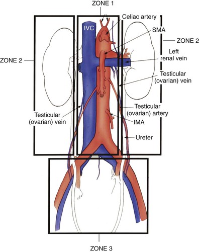
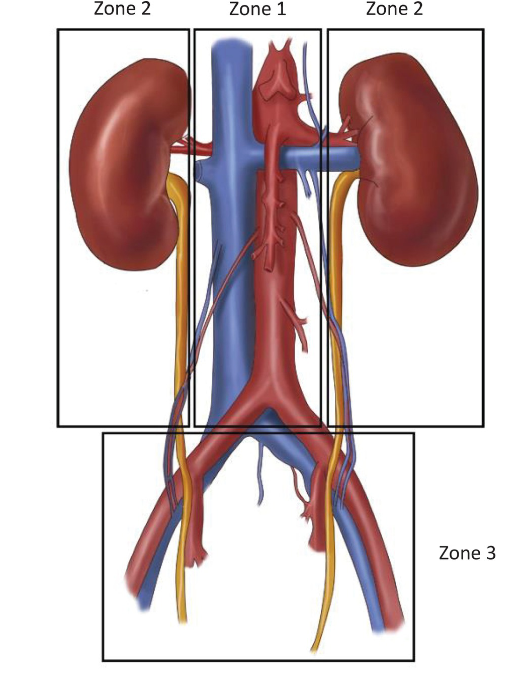
Vascular Trauma Thoracic Key
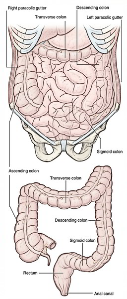
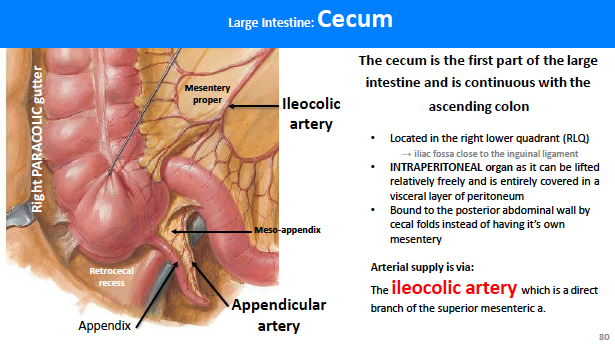
Easy Notes On Colon Learn In Just 4 Minutes Earth S Lab

Abdomen Flashcards Quizlet
Lab 23 Structures Lab 23 Structures Flashcards Memorang
Peritoneum Dr Mehul Tandel
Studying The Peritoneum With Human Anatomy Atlas 2020

The Colon Ascending Transverse Descending Sigmoid Teachmeanatomy

Pin By Dharmin Trivedi On Anatomy Anatomy Cavities Fluid

Image Result For Peritoneogram Radiology Radiology Medical Image
Https Encrypted Tbn0 Gstatic Com Images Q Tbn 3aand9gcshgccvhjwl4fga6xmtvrtmvxwjd3da6xh699xbs0yifrpvajgq Usqp Cau

Lymph Vessels And Nodes Of Small Intestine Anatomy Thoracic Duct Cisterna Chyli Intestinal Lymphatic Tr Intestines Anatomy Lymph Vessels Thoracic Duct
Epos
Https Jumed14 Weebly Com Uploads 5 8 7 5 58753271 Sheet8 Pdf
Https Link Springer Com Content Pdf 10 1007 2f0 387 21578 6 12 Pdf

Abdominal Wall And Hernias Abdominal Key

Surface Anatomy Vessels Muscles And Peritoneum Ppt Download
Colon Anatomy

Figure 4 3 From Open Abdomen Semantic Scholar
Abdominal Cavity 1 Online Presentation

Abdominal Wall And Cavity Flashcards Quizlet
Sectional Anatomy Of Abdomen

Netter Atlas Of Human Anatomy 7th Ed 2018 Hotrotailieuykhoa Blogspot Com Flip Book Pages 501 550 Pubhtml5
Https Nanopdf Com Download 12abdominal Fascia Pdf

1 Short Descending Mesocolon And Redundant Sigmoid Colon In The Right Download Scientific Diagram
Pelvis Cross Section Anatomy
Abdominal Aorta And Splachnic Vessels Chapter 31 Atlas Of Surgical Techniques In Trauma

Ultrasound In Preoperative Assessment Of Pelvic And Abdominal Spread In Patients With Ovarian Cancer A Prospective Study Fischerova 2017 Ultrasound In Obstetrics Amp Gynecology Wiley Online Library

Ppt Fascia Of The Abdomen Powerpoint Presentation Free Download Id 1978915

Lateroconal Fascia Radiology Reference Article Radiopaedia Org

Radiology Quiz 29199 Radiopaedia Orgviewing Playlist 3 005 Ascites And Peritoneal Fluid Collection Radiopaedia Org
Peritoneum
Anatomy Exam 3 Exam 3 Flashcards Memorang

29 Pelvis Radiology Key